Ankle Open Reduction Internal Fixation (ORIF)
An Ankle ORIF is an orthopaedic surgical procedure to realign and stabilise fractured bones in the ankle using plates, screws, and/or rods.
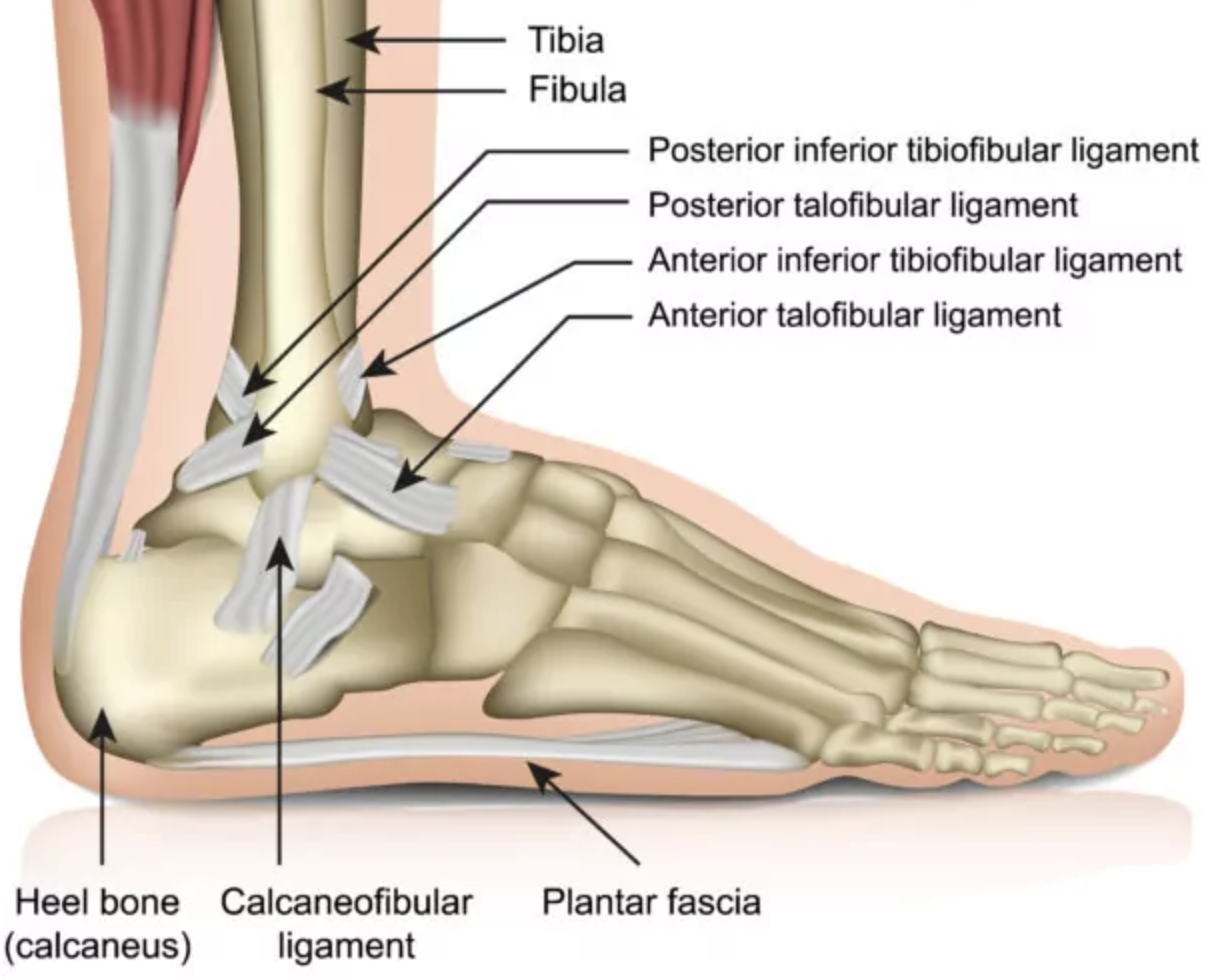
Relevant Anatomy
The ankle joint involves the distal tibia, fibula, talus, and associated ligaments. Familiarity with medial, lateral, and posterior malleolus anatomy is crucial.
1. Instruments and Equipment Checklist
Basic Ortho Tray (Sterile)
- Scalpel blades (No. 10, 15)
- Mayo scissors (curved and straight)
- Metzenbaum scissors
- Tissue forceps (toothed, Adson, DeBakey)
- Needle holders
- Periosteal elevator (Freer or Key)
- Bone holding forceps
- Retractors (Senn, Army-Navy, Hohmann)
- Electrocautery (bovie)
- Power drill with battery
- Suction (Yankauer or Frazier)
Specialised Instruments
- Orthopaedic screw set (cortical/cancellous)
- Plating system (locking/non-locking)
- Depth gauge
- Drill bits (2.0 mm, 2.5 mm, 3.2 mm)
- Taps and screwdrivers
- Reduction clamps
- K-wires (for temporary fixation)
Sutures
- 3-0 and 4-0 Monocryl or Vicryl
- 2-0 Nylon or staples for skin closure
Other Equipment
- Tourniquet and control unit
- Leg holder or bean bag
- Positioning foam or gel pads
- Pulse lavage for wound irrigation
- Sterile dressing supplies
- Foot drapes
Fluids and Medications
- Antibiotics: Cefazolin IV pre-op per protocol
- Normal saline: For irrigation and wound washout
- Local anaesthetic: Bupivacaine or lignocaine with adrenaline (surgeon preference)
- Tranexamic acid (TXA): May be used to reduce bleeding
2. Before Knife to Skin
- Check and lay out all instruments on scrub table
- Initial count of swabs, instruments, and sharps
- Apply tourniquet but do not inflate until requested
- Confirm implant trays and sizes are available and opened sterilely
- Coordinate with radiographer for intra-op imaging
Prepping and Draping
- Prep foot, ankle, and leg up to knee using alcoholic chlorhexidine
- Use leg holder or beanbag for support
- Drape with foot drapes or a full extremity pack
- Ensure tourniquet lines and bovie cables are kept sterile
3. Intraoperative Stages
- Incision and Exposure: Medial or lateral approach depending on fracture type; retractors in place.
- Fracture Reduction: Use clamps, K-wires, or manual reduction techniques; confirm with imaging.
- Internal Fixation: Drill, tap, measure, insert screws and plates in correct sequence.
- Check Alignment: Use C-arm to confirm fracture reduction and hardware position.
- Irrigation: Pulse lavage the wound thoroughly.
- Closure: Layered closure with absorbable sutures and skin closure with staples or nylon.
4. Post-Op Tasks
- Final count of swabs, sharps, and instruments
- Apply sterile dressing and padding
- Assist with tourniquet deflation and check for bleeding
- Clean and pack away instruments
- Complete intraoperative documentation
- Hand over to PACU nurse with relevant details and implants used