Lymph Node Dissection
A Lymph Node Dissection is a procedure to remove lymph nodes and surrounding soft tissue from levels III, IV, and occasionally V of the neck, often for staging or treatment of head and neck malignancies.
Relevant Anatomy
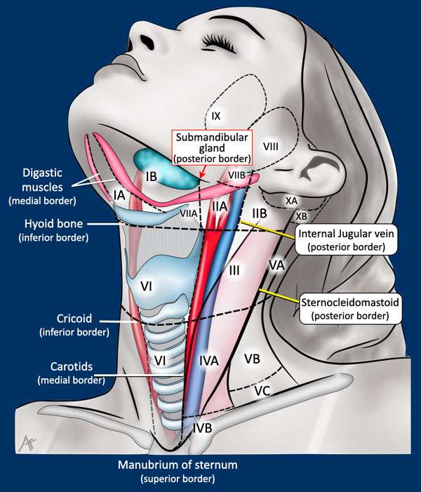
The procedure involves careful dissection near vital structures such as the internal jugular vein (IJV), spinal accessory nerve (SAN), sternocleidomastoid muscle, vagus nerve, and carotid artery. Lymph node levels III and IV are frequently targeted.
1. Instruments and Equipment Checklist
Basic ENT Tray (Sterile)
- Scalpel blades (No. 10, 15)
- Mayo scissors (curved and straight)
- Metzenbaum scissors
- Tissue forceps (toothed, Adson, DeBakey)
- Needle holders
- Hemostats (mosquito, Kelly, Crile)
- Retractors (skin hooks, rake retractors)
- Electrocautery (bovie)
- Suction (Frazier or Yankauer)
Specialised Instruments
- Silastic vessel loops
- Clip applier with Hemoclips
- Nerve stimulator (if used)
- Dissectors (tonsil, peanut)
- Skin stapler (optional)
Sutures
- 3-0 and 4-0 Vicryl or Monocryl for deep layers
- 3-0 Nylon or staples for skin closure
Other Equipment
- Closed suction drain (e.g. Jackson-Pratt)
- Head ring or foam donut for head support
- Neck roll for patient positioning
- Pulse lavage (optional for irrigation)
- Sterile dressing supplies
- ENT head-and-neck drape pack
Fluids and Medications
- Antibiotics: Cefazolin IV pre-op per protocol
- Normal saline: For irrigation
- Local anaesthetic: Lignocaine with adrenaline (field block)
- Steroids: May be given intra-op to reduce edema
2. Before Knife to Skin
- Position patient supine with neck extended using a roll
- Initial count of swabs, sharps, and instruments
- Ensure suction, light source, and electrocautery are functional
- Confirm drain, vessel loops, and clip appliers are present
- Verify correct side and levels of dissection with surgical team
Prepping and Draping
- Prep entire neck and upper chest using alcoholic chlorhexidine
- Drape with ENT-specific pack ensuring full exposure of lower neck
- Secure bovie, suction, and light cables safely and sterilely
3. Intraoperative Stages
- Incision and Exposure: Transverse incision along skin crease in lower neck; raise subplatysmal flaps
- Identification of Landmarks: Expose IJV, SAN, carotid sheath; apply vessel loops as needed
- Dissection: Systematic removal of nodal tissue from levels III and IV, avoiding neurovascular damage
- Hemostasis: Meticulously cauterise or clip bleeding vessels
- Irrigation: Use saline to wash surgical field
- Closure: Place drain, close platysma and skin using appropriate suture or staples
4. Post-Op Tasks
- Final count of swabs, sharps, and instruments
- Secure and label drain with suction applied
- Apply sterile dressing over wound and drain site
- Assist with head repositioning and monitor airway patency
- Clean and return instruments for reprocessing
- Document drain output plan, anatomical structures preserved, and handover to PACU team